File:Didelphidae early development of blastoderm.jpg

Size of this preview: 795 × 599 pixels. Other resolutions: 318 × 240 pixels | 637 × 480 pixels | 1,019 × 768 pixels | 1,045 × 788 pixels.
Original file (1,045 × 788 pixels, file size: 621 KB, MIME type: image/jpeg)
File information
Structured data
Captions
Captions
Add a one-line explanation of what this file represents
Summary
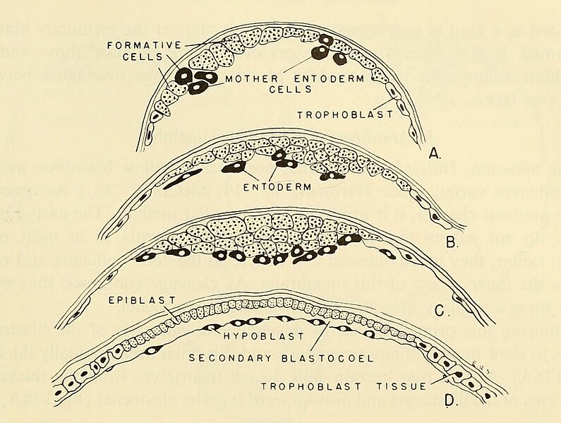
edit| DescriptionDidelphidae early development of blastoderm.jpg | Fig. 176. Early development of blastoderm of the opossum. (Modified from Hartman, '16.) (A) Blastocyst wall composed of one layer of cells from which entoderm ceils are migrating inward. (B-D) Later development of the formative portion of the blastoderm. Two layers of cells are present in the formative area, viz., an upper epiblast layer and a lower hypoblast. Trophoblast cells are shown at the margins of the epiblast and hypoblast layers. |
| Date | |
| Source | https://archive.org/details/comparativeembry00nels/page/363/mode/1up?view=theater&q=BLASTULATION+ Comparative embryology of the vertebrates; with 2057 drawings and photos. grouped as 380 illustrations. |
| Author | Nelsen, Olin E. |
Licensing
edit| Public domainPublic domainfalsefalse |
|
This work is in the public domain in its country of origin and other countries and areas where the copyright term is the author's life plus 70 years or fewer. Note that a few countries have copyright terms longer than 70 years: Mexico has 100 years, Jamaica has 95 years, Colombia has 80 years, and Guatemala and Samoa have 75 years. This image may not be in the public domain in these countries, which moreover do not implement the rule of the shorter term. Honduras has a general copyright term of 75 years, but it does implement the rule of the shorter term. Copyright may extend on works created by French who died for France in World War II (more information), Russians who served in the Eastern Front of World War II (known as the Great Patriotic War in Russia) and posthumously rehabilitated victims of Soviet repressions (more information).
| |
| This file has been identified as being free of known restrictions under copyright law, including all related and neighboring rights. | |
https://creativecommons.org/publicdomain/mark/1.0/PDMCreative Commons Public Domain Mark 1.0falsefalse

|
This file, which was originally posted to an external website, has not yet been reviewed by an administrator or reviewer to confirm that the above license is valid. See Category:License review needed for further instructions.
|
File history
Click on a date/time to view the file as it appeared at that time.
| Date/Time | Thumbnail | Dimensions | User | Comment | |
|---|---|---|---|---|---|
| current | 01:47, 12 March 2024 |  | 1,045 × 788 (621 KB) | Rasbak (talk | contribs) | {{Information |description=Fig. 176. Early development of blastoderm of the opossum. (Modified from Hartman, '16.) (A) Blastocyst wall composed of one layer of cells from which entoderm ceils are migrating inward. (B-D) Later development of the formative portion of the blastoderm. Two layers of cells are present in the formative area, viz., an upper epiblast layer and a lower hypoblast. Trophoblast cells are shown at the margins of the epiblast and hypoblast layers. |date=1953 |source=https:/... |
You cannot overwrite this file.
File usage on Commons
There are no pages that use this file.